GAFAMS, STARTUPS & INNOVATION IN HEALTHCARE by PHARMAGEEK
117.5K views | +30 today
Follow
GAFAMS, STARTUPS & INNOVATION IN HEALTHCARE by PHARMAGEEK
Your new post is loading...
Rescooped by Lionel Reichardt / le Pharmageek from healthcare technology
Scoop.it!

Grant awarded to develop artificial intelligence to improve stroke screening and treatment in smaller hospitals

Grant awarded to develop artificial intelligence to improve stroke screening and treatment in smaller hospitals | GAFAMS, STARTUPS & INNOVATION IN HEALTHCARE by PHARMAGEEK | Scoop.it

New artificial intelligence technology that uses a common CT angiography (CTA), as opposed to the more advanced imaging normally required to help identify patients who could benefit from endovascular stroke therapy (EST), is being developed at The University of Texas Health Science Center at Houston (UTHealth).


Two UTHealth researchers worked together to create a machine-learning artificial intelligence tool that could be used for assessing a stroke at every hospital that takes care of stroke patients - not just at large academic hospitals in major cities. 


Research to further develop and test the technology tool is funded through a five-year, $2.5 million grant from the National Institutes of Health (NIH). 


"The vast majority of stroke patients don't show up at large hospitals, but in those smaller regional facilities. And most of the emphasis on screening techniques is only focused on the technologies used in those large academic centers. With this technology, we are looking to change that," said Sunil Sheth, MD, assistant professor of neurology at McGovern Medical School at UTHealth.


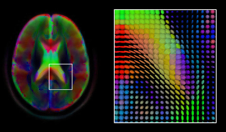
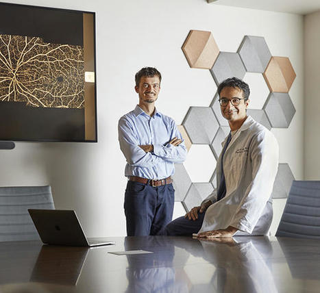
Sheth set out with Luca Giancardo, PhD, assistant professor with the Center for Precision Health at UTHealth School of Biomedical Informatics, to develop a quicker way to assess patients. The result was a novel deep neural network architecture that leverages brain symmetry. Using CTAs, which are more widely available, the system can determine the presence or absence of a large vessel occlusion and whether the amount of "at-risk" tissue is above or below the thresholds seen in those patients who benefitted from EST in the clinical trials.


"This is the first time a data set is being specifically collected aiming to address the lack of quality imaging available for stroke patients at smaller hospitals," Giancardo said.


read the complete press release with further details on the work at https://www.uth.edu/news/story.htm?id=9fccdefb-ff91-4775-a759-a786689956ea


Via nrip
No comment yet.
Rescooped by Lionel Reichardt / le Pharmageek from healthcare technology
Scoop.it!

Algorithms begin to show practical use in diagnostic imaging  #esante #hcsmeufr

Algorithms begin to show practical use in diagnostic imaging  #esante #hcsmeufr | GAFAMS, STARTUPS & INNOVATION IN HEALTHCARE by PHARMAGEEK | Scoop.it

Algorithms based on machine learning and deep learning, intended for use in diagnostic imaging, are moving into the commercial pipeline.


However, providers will have to overcome multiple challenges to incorporate these tools into daily clinical workflows in radiology.


There now are numerous algorithms in various stages of development and in the FDA approval process, and experts believe that there could eventually be hundreds or even thousands of AI-based apps to improve the quality and efficiency of radiology.


The emerging applications based on machine learning and deep learning primarily involve algorithms to automate such processes in radiology as detecting abnormal structures in images, such as cancerous lesions and nodules. The technology can be used on a variety of modalities, such as CT scans and X-rays. The goal is to help radiologists more effectively detect and track the progression of diseases, giving them tools to enhance speed and accuracy, thus improving quality and reducing costs.


While the number of organizations incorporating these products into daily workflows is small today, experts expect many providers to adopt these solutions as the industry overcomes implementation challenges.


Data dump
Radiologists’ growing appreciation for AI may result from the technology’s promise to help the profession cope with an explosion in the amount of data for each patient case.


Radiologists also are grappling with the growth in data from sources outside radiology, such as lab tests or electronic medical records. This is another area where AI could help radiologists by analyzing data from disparate sources and pulling out key pieces of information for each case,.


There are other issues that AI could address as well, such as “observer fatigue,” which is an “aspect of radiology practice and a particular issue in screening examinations where the likelihood of finding a true positive is low,” wrote researchers from Massachusetts General Hospital and Harvard Medical School in a 2018 article in the Journal of the American College of Radiology.


These researchers foresee the utility of an AI program that could identify cases from routine screening exams with a likely positive result and prioritize those cases for radiologists’ attention.

AI software also could help radiologists improve worklists of cases in which referring physicians already suspect that a medical problem exists.


read more at the original source: https://www.healthdatamanagement.com/news/algorithms-begin-to-show-practical-use-in-diagnostic-imaging


Via nrip
No comment yet.